Give your patients your best work
In surgical oncology, such as breast conserving surgery, rapid assessment of the excised specimen is of utmost importance. You want to be confident that your surgery reached its goal of resecting the target lesion, so that your patients recovery can be promoted.
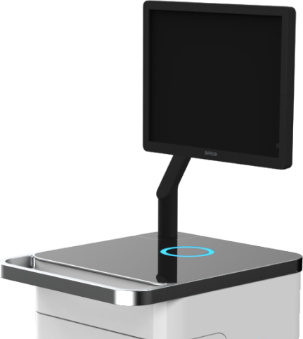
The first PET-CT specimen imager for the OR
Aura 10 has made molecular imaging available in the OR for the first time. As a result, surgeons and imaging specialists have never been able to perform specimen imaging with so much confidence. Ultimately, your patients will be the ones who benefit.
Benefits of intra-operative specimen imaging
Less chance of resurgery
The AURA 10 combines the picomolar sensitivity of PET with high-precision 3D image reconstruction. This gives you all the imaging capabilities you need to image the targeted tissue in a single procedure, to close your patient with more confidence, and to reduce the probability of resurgery.
More peace of mind for your patients
Using the best available intraoperative imaging technology will significantly decrease the anxiety level of the patient. This stress reduction will contribute to your patient’s recovery.
Reduced anesthesia time
A more streamlined workflow and real-time collaboration with your radiology department can shorten the procedure. This may also reduce anesthesia time for your patient and decrease the risk of postoperative complications.
Better cosmetic outcome
Intraoperative imaging can help you to remove less tissue in the initial surgery and to obtain a better cosmetic result.