Imaging Case 5:
Prostate Cancer - Adenocarcinoma
Radical prostatectomy often requires sacrificing the neurovascular bundle, which impacts the quality of life of prostate cancer patients. A nerve-sparing approach can be used, but this involves an increased risk of incomplete resection. Therefore, it is interesting to know intraoperatively whether the tumor has been sufficiently removed when performing nerve-sparing surgery.
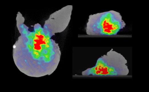
Prostate cancer cells have an increased PSMA expression, which is how they can be distinguished from healthy prostatic tissue on whole-body PSMA PET-CT.[1] In this clinical case, we highlight how intraoperative specimen PSMA PET-CT imaging visualizes the tumor within the specimen. This could aid the surgeon in deciding whether it is necessary to remove more tissue intraoperatively when aiming for a complete resection.
This clinical case is courtesy of Dr. C. Darr and Prof. B. Hadaschik, Department of Urology, Universitätsmedizin Essen.
[1] Okarvi, S. M. Recent developments of prostate-specific membrane antigen (PSMA)-specific radiopharmaceuticals for precise imaging and therapy of prostate cancer: an overview. Clin. Transl. Imaging 7, 189–208. issn: 22817565 (June 2019).