Imaging Case 9:
Papillary thyroid carcinoma
Papillary thyroid carcinoma is the most common type of thyroid cancer. Depending on the stage at diagnosis, it is treated with a combination of (total) thyroidectomy, cervical lymphadenectomy and/or radioiodine treatment[1]. If a lymph node metastasis is detected at follow-up, resection of the invaded lymph nodes can be considered. However, confirming intraoperatively that the target lymph nodes have been resected can be a challenge.
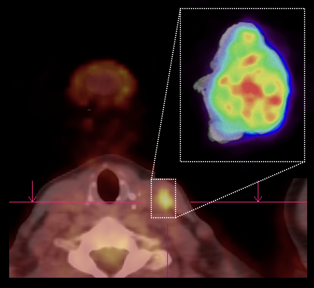
In this clinical case we show how a detailed view of resected lymph nodes can be obtained with high-resolution specimen PET-CT imaging. This information can help the surgeon determine if the targeted lymph nodes have been resected right at the point of surgery. This case is presented with the support of dr. Valérie Vergucht and prof. dr. Bieke Lambert of AZ Maria Middelares, Ghent, Belgium.