Story
Invasive ductal carcinoma
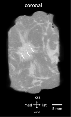
A 65-year-old woman with invasive ductal carcinoma underwent breast-conserving surgery followed by sentinel lymph node resection.
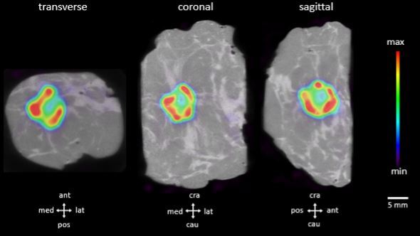
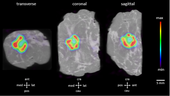
The resected breast specimen was imaged immediately after resection.
Microcalcifications can be detected on the CT specimen images. Figure 2 depicts a coronal section of the CT scan. The white arrows indicate the microcalcifications in the resected breast tumor.