Imaging Case 1:
Breast - Invasive ductal carcinoma
A 65-year-old woman with invasive ductal carcinoma underwent breast-conserving surgery followed by sentinel lymph node resection.
 Images and case information courtesy of dr. Menekse Göker, Department of Gynaecology, Ghent University Hospital.”
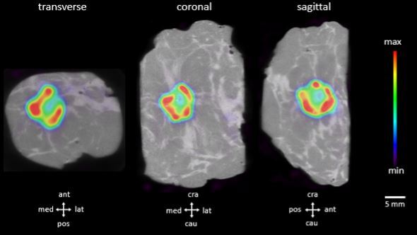
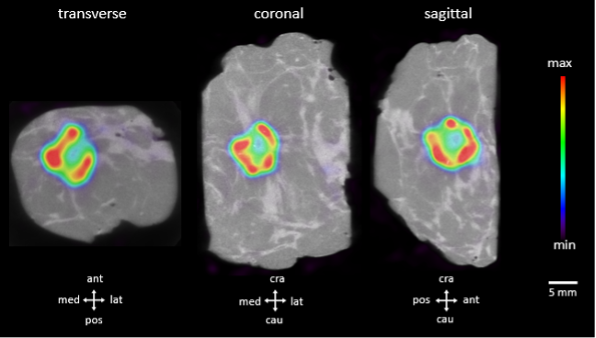
The patient was pre-operatively injected with 18F-FDG at the nuclear medicine department. The resected breast specimen was imaged immediately after resection. Figure 1 shows three orthogonal views of the PET-CT specimen images. The PET images are represented in color scale superimposed on the CT images in greyscale. The breast tumor is visible in the PET-CT specimen images as the large bright colored region at the center of the specimen.
Figure 1 shows the transverse, coronal and sagittal slides of the PET-CT specimen scan. The invasive tumor is represented by a bright colorful region at the center of the specimen. Surgical orientation of the specimen images is shown at the bottom of each section. The PET tracer scale bar is depicted at the right hand side.
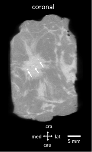
Microcalcifications can be detected on the CT specimen images. Figure 2 depicts a coronal section of the CT scan. The white arrows indicate the microcalcifications in the resected breast tumor. In addition to the information provided by the PET image, this may also allow confirming that all microcalcifications were resected.
Figure 2 shows the coronal view of the CT scan of the resected breast tumor. Microcalcifications can be clearly detected on the CT images (white arrows).
Do you want to discover more on this case? You can download the complete case study using the button below.