XEOS AURA 10 PET-CT installed at Universitätsmedizin Essen
UK Essen is the first prostate cancer center with access to intraoperative specimen PET-CT imaging. The information from this new imaging modality could be used to enhance surgery, improve outcomes and give patients and surgeons peace of mind.
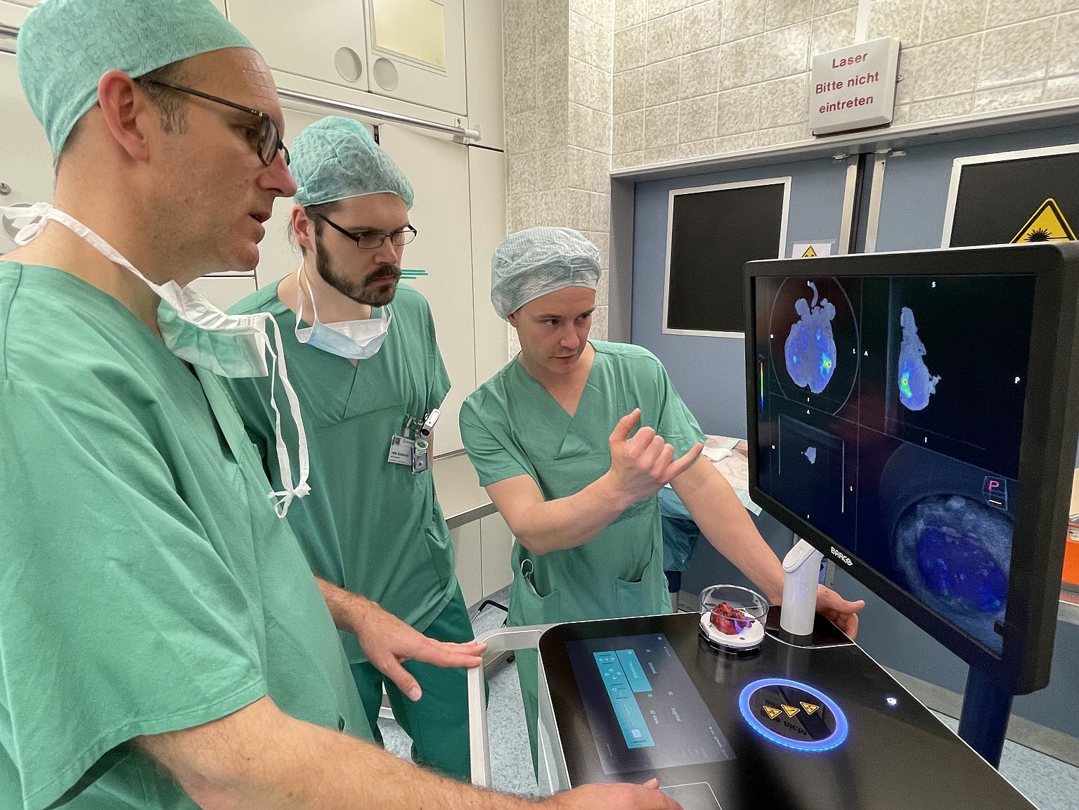
The AURA 10 is the first-ever specimen PET-CT imager, designed for use in the operating room. Specimen PET-CT imaging offers surgeons an immediate view of the resected tissue specimen during the surgical procedure. The images enable the surgeon to assess intraoperatively with more confidence whether the targeted tissue – such as prostate cancer – was removed. This can aid in enhancing surgical outcomes.
The XEOS AURA 10 was installed at the Department of Urology at UK Essen. Together with the Department of Nuclear Medicine, there is already plenty experience with novel imaging techniques, both preclinically and clinically. With this install, UK Essen becomes the first prostate cancer center with access to intraoperative specimen PET-CT imaging. “We are excited to see the commitment of UK Essen to innovate in intraoperative imaging,” says Roel Van Holen, CEO of XEOS. “We are convinced that the combination of clinical and scientific excellence in this team is ideal to foster the further exploration of the added value of specimen PET-CT imaging.”
Improving prostate cancer treatment
In prostate cancer treatment, the goal of intraoperative specimen imaging is to support surgeons to perform nerve-sparing procedures with more confidence. “Our first experience shows that specimen PET-CT imaging brings a wealth of interesting information to the operating room,” says Prof. Boris Hadaschik, Chair of the Department of Urology. “The installation of the device now, allows us to expand our possibilities – in prostate cancer and in other malignancies. Our goal is to improve patient outcomes by enhancing surgery.”
The Department of Nuclear Medicine is also an important partner in the implementation of specimen PET-CT imaging at UK Essen. Prof. Ken Herrmann, Chair of the Department of Nuclear Medicine, also sees possibilities beyond Urology: “As the AURA 10 can image any PET radiotracer, this technology could also help surgeons in other therapeutic areas where PET imaging is currently standard of care for diagnosis and follow-up.”
About XEOS
Founded in 2019 in Ghent, Belgium, XEOS is an expert in specimen imaging, and focuses on improving outcomes in surgery through innovations in intraoperative imaging. The XEOS team is passionate about expanding the use of molecular imaging to optimize clinical workflows and improve patient outcomes. The company has an admirable track record of in-house PET & CT design. XEOS is an ISO 13485 certified company.
For more information on XEOS please contact:
Ms. Natacha Sweert
natacha.sweert@xeos.care
+32 9 277 77 94 (Belgium)
www.xeos.care
About the prostate cancer center at University Medical Center Essen
In localized high-risk prostate cancer, PSMA-based molecular diagnostics demonstrated a significantly higher accuracy compared to conventional imaging, resulting in an increasing use in staging prior to surgery. As a certified prostate cancer center, the Departments of Urology and Nuclear Medicine at the University Medical Center Essen strive to continuously improve local treatment options. The possibility to transfer molecular diagnostics into the intraoperative field is of great scientific interest and has the potential to further improve treatment of our patients in the future.
For more information about the prostate cancer center at University Medical Center Essen please contact:
Mr. Burkhard Büscher
burkhard.buescher@uk-essen.de
+49 201 723 2115 (Germany)
https://urologie.uk-essen.de/