Imaging Case 24:
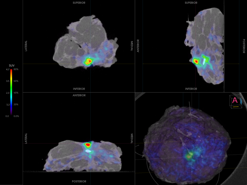
Head & Neck cancer - Squamous Cell Carcinoma of the palate and alveolus
Surgeons treating oral squamous cell carcinoma aim for a minimum marginal clearance of 5 mm. The complex three-dimensional anatomy of the oral cavity makes achieving these clear resection margins particularly challenging. When margins are close or involved, patients often require re-excision. However, histopathology analysis of margins takes at least fourteen days, making additional resection impractical once final results are available. As a result, adjuvant radiotherapy is commonly required, increasing morbidity—sometimes with lifelong consequences. Moreover, involved margins significantly raise the risk of treatment failure, particularly when a bone margin is affected, as adjuvant therapy is less effective in bone than in soft tissue.
This case highlights the potential of intraoperative specimen PET-CT imaging to address these challenges by providing real-time visualization of the tumor within the resected specimen. By enabling immediate, informed decision-making during surgery, this technique may improve resection completeness and reduce the need for additional treatment.
The case is presented with the support of Mr. Gary Walton, consultant head and neck surgeon, Dr. Oludolapo Adesanya, consultant radiologist, and colleagues of University Hospitals Coventry & Warwickshire, Coventry, United Kingdom, as part of the ‘eXcision’ trial (IRAS ID: 342171, REC reference 24/YH/0137). The ‘eXcision’ trial is a single center prospective pilot study investigating the diagnostic performance of high-resolution specimen PET-CT in prostate cancer and head and neck cancer resection.
Read the whole story


