Imaging Case 12:
Breast cancer – Invasive Lobular Carcinoma
During breast-conserving surgery it is common practice to perform intraoperative assessment of the resected tumor specimen to assess if the resection was successful. This is especially useful for patients diagnosed with invasive lobular carcinoma, as the positive margin rate is higher in this population. The most frequently used techniques such as specimen radiography or frozen section assessment all have their drawbacks regarding performance and/or efficiency.
In this clinical case, which is part of the multi-center prospective BrIMA study (NCT04999917), we highlight how intraoperative specimen PET-CT imaging can bring more confidence into the operating room. This gives the surgeon a clear view of what was resected – and the ability to act upon this information at the point of surgery.
Patient History
A 64-year-old woman with invasive lobular carcinoma underwent breast-conserving surgery followed by sentinel node resection. The tumor size on preoperative imaging was 54mm, biopsy showed a grade 2 tumor with receptor status ER+/PR+/HER2+. Preoperative staging was cT3N0M0.
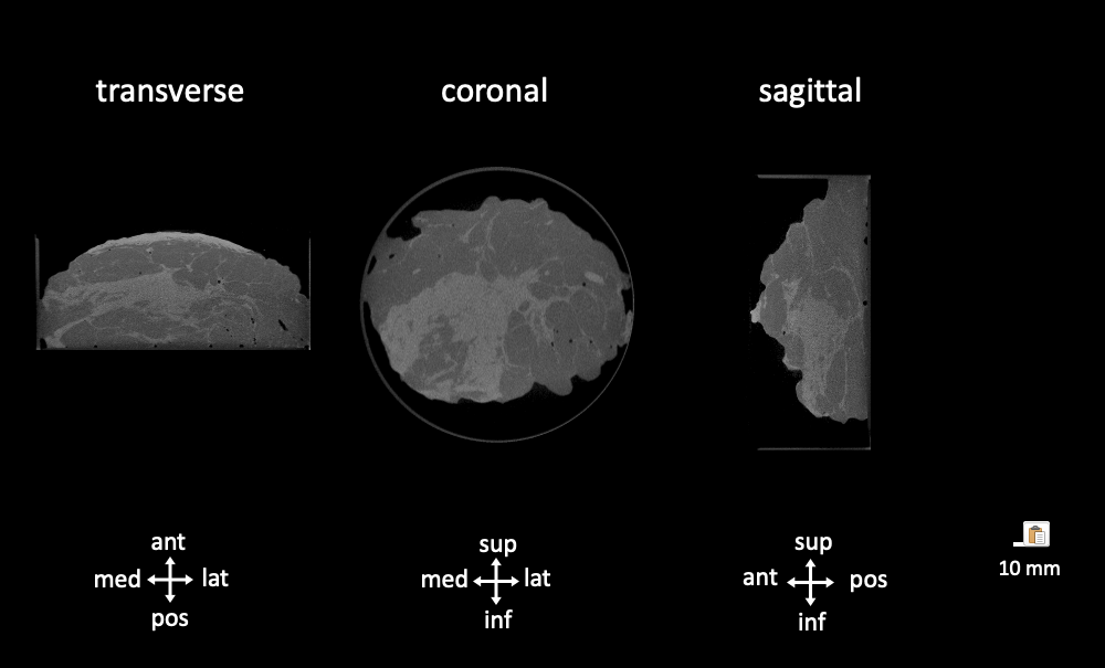 Figure 1. Transverse, coronal and sagittal slices of the CT specimen images of an invasive lobular carcinoma. A dense lump is visualized slightly excentric in the specimen towards the inferomedial border of the specimen. Abbreviations: med, medial; lat, lateral; sup, superior; inf, inferior.
PET-CT specimen images
For sentinel node localization, the patient received an intratumoral injection with 152 MBq of 99mTc on the morning of surgery at the nuclear medicine department. An intravenous injection with 68 MBq (0.80 MBq/kg) of 18F-FDG was also given at the nuclear medicine department. The patient was then transferred to the operating theatre and breast-conserving surgery was performed per standard protocol.
The resected breast specimen was imaged immediately after resection and the images were interpreted by the surgeon in the operating room. This was approximately 160 min after 18F-FDG injection.
The CT (figure 1) visualizes the entire specimen in 3D and shows a dense lump in the specimen, localized slightly towards the inferomedial side of the specimen. This already provides an indication of where the tumor is located in the specimen.
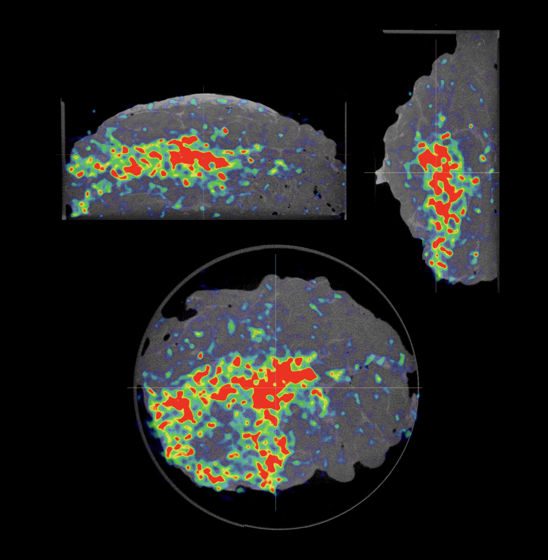
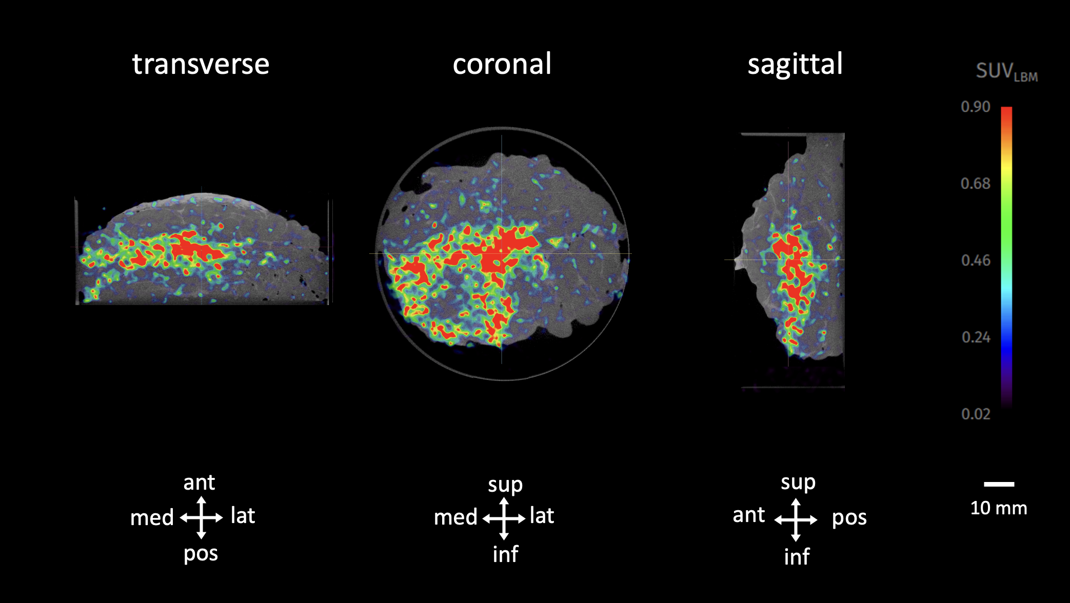 Figure 2. Transverse, coronal and sagittal slices of the PET-CT specimen images of an invasive lobular carcinoma. The 18F-FDG uptake is visible in the color overlay and reaches the inferomedial border of the specimen. Final histopathology confirmed a positive surgical margin. Specimen orientation is as indicated. Abbreviations: med, medial; lat, lateral; sup, superior; inf, inferior.
Fig. 2 shows the combined PET-CT images, highlighting areas of interest inside the specimen. The colored overlay demonstrates increased 18F-FDG uptake, which indicates metabolically more active regions such as invasive cancer cells.
In this case, 18F-FDG uptake reaches the inferomedial border of the imaged specimen. Based on this image, the surgeon decided to take a cavity shave in this area. Postoperative final pathology showed that there was indeed a positive surgical resection margin in this area.
Histopathological Evaluation
Immediately after specimen imaging, the resected specimen was sent to the pathology department for routine histopathological evaluation, which was available after several days. Table 1 shows the results of histopathology. The additional cavity shave yielded a final negative margin status. The patient could therefore immediately continue postoperative therapy, without the need for a reoperation.
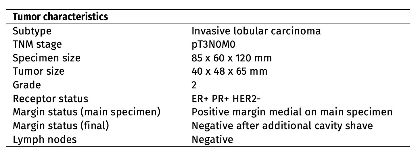 Table 1. A summary of the histopathological findings of the resected specimens.
Discussion and conclusion
In this interesting case, the surgeon decided to perform an additional cavity shave based on specimen PET-CT imaging as 18F-FDG uptake was reaching the inferomedial border of the specimen. Histopathological evaluation also showed tumor cells reaching the border of the specimen in this area. In this patient this additional cavity shave helped to avoid the need for reoperation and the associated burden.
REFERENCES