Imaging Case 2:
Breast - Invasive lobular carcinoma
An 84-year-old woman was diagnosed with an invasive lobular carcinoma. Breast-conserving surgery including lumpectomy and sentinel lymph node resection was performed.
 Images and case information courtesy of dr. Menekse Göker, Department of Gynaecology, Ghent University Hospital.”
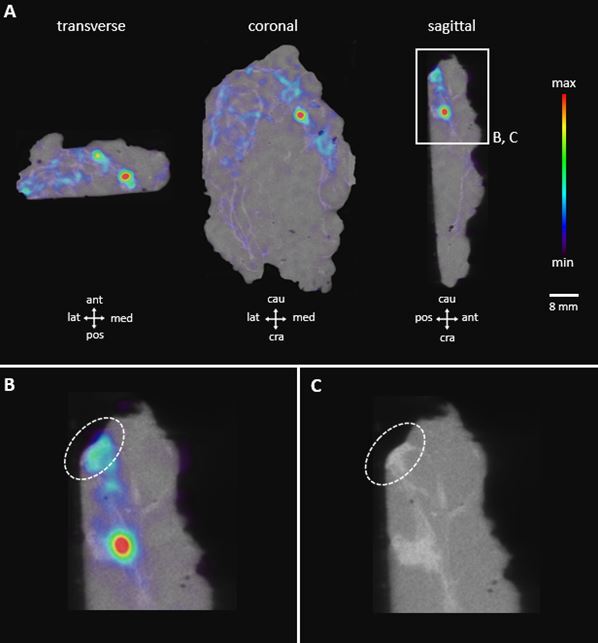
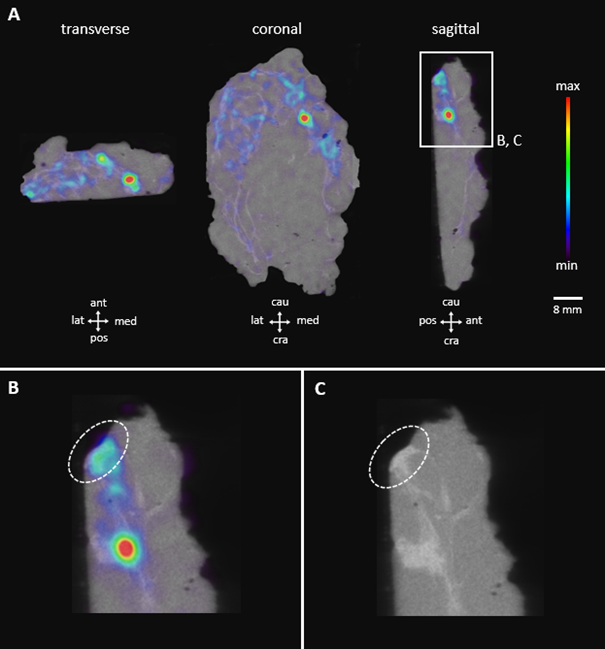
The patient was pre-operatively injected with 18F-FDG (IV) at the nuclear medicine department. Figure 1A shows three orthogonal slices of the specimen PET-CT image. Increased 18F-FDG uptake is visualized (colored hotspots) which indicates metabolically more active cells such as the invasive lobular carcinoma cells. There is an area of increased FDG uptake close to the top of the sagittal section (caudal margin). An enlargement of this suspicious area is provided in Figure 1B (PET-CT) and Figure 1C (CT only). The 18F-FDG uptake is represented by a bright colorful region that reaches the specimen border (Figure 1B, dashed ellipse). The CT image confirms that dense tissue is also reaching the border (Figure 1C, dashed ellipse)*.
Figure 1 represents three orthogonal views (A) of the PET-CT specimen images. The invasive lobular carcinoma is represented by the 18F-FDG hotspots that are reaching the tissue borders delineated by the CT image. The surgical orientation of the specimen is indicated at the bottom of each view. The color bar at the right shows the relative 18F-FDG activity in the specimen. Figure 1B (PET CT) and Figure 1C (CT only) show an enlargement of the suspicious margin observed at the top of the sagittal section. The suspicious margin is indicated by the dashed ellipse.
The histopathological analysis confirmed a positive margin (i.e. involvement of tumor cells at the tissue border) with a length of 2 mm at the caudal margin. This is also supported by the findings of radiotracer uptake reaching the caudal border of the specimen on the PET-CT image. After a multidisciplinary consultation, it was decided to schedule a mastectomy.
Do you want to discover more on this case? You can download the complete case study using the button below.