Imaging Case 25:
Breast cancer - Invasive Ductal Carcinoma
Over the past 30 years, breast-conserving surgery (BCS) with radiation therapy has become the standard treatment for early-stage breast cancer, offering survival rates comparable to mastectomy with better cosmetic outcomes.
Successful BCS requires complete tumor removal with negative margins, as positive margins increase recurrence risk and often lead to reoperation. Therefore, specimen imaging, supporting margin assessment, is vital in breast cancer surgery. Intraoperative margin assessment helps ensure clear margins, with methods like specimen radiography and frozen section analysis. However, each of these methods have limitations in accuracy, efficiency, or practicality [1].
Recent publications have shown that high-resolution specimen PET-CT imaging with FDG-based radiotracers can provide an accurate view of the resected breast tumor specimen and may help enable assessing completeness of resection in a fast and efficient way [2,3]. In this clinical case, we illustrate how intraoperative specimen PET-CT imaging provides a precise, real-time visualization of resected tissue, allowing the surgeon to make immediate, well-informed decisions and optimize surgical outcomes.
This case is presented with the support of Z Maria Middelares, Ghent, Belgium, and is part of the investigator driven study to explore potential indications of an intraoperative specimen PET-CT imager (Trial registration number: BUN: B0172022000009).

Patient history
A 70-year-old woman, with a history of surgery on her left breast for a benign lesion, was diagnosed with breast cancer in the same region through ultrasound and mammography. Imaging revealed a lesion (diameter approximately 30 mm) without signs of lymph node invasion. Biopsy showed a grade 2 Invasive Ductal Carcinoma (IDC), NST, with receptor status positive (ER, PR, HER2) and Ki-67 index <10%. Clinical staging was determined as T2N0M0. Based on these findings, the patient was scheduled for breast-conserving surgery (BCS) with sentinel node removal.
Figure 1: Transverse, coronal, and sagittal slices of the specimen PET-CT images of the tumor specimen, together with a 3D view. Specimen orientation is as indicated. The PET tracer scale bar is depicted on the left-hand side. The image clearly outlined the skin, for which the 18F-FDG uptake extended to this area. Across the entire specimen, uptake did not reach the border of the tissue.
Specimen PET-CT imaging
The patient was intravenously injected with 0.8 MBq/kg of 18F-FDG 60 minutes prior to surgery at the Nuclear Medicine Department. The patient was then transferred to the operating theatre and breast-conserving surgery was performed.
During the procedure, the surgeon palpated and excised the tumor. Initially, the goal was to preserve the skin. However, as the tumor was palpable up to the skin border, the surgeon decided to dissect the portion of skin overlaying the tumor.
The tumor specimen was resected and scanned 141 minutes post-injection using the specimen PET-CT imager, see Fig. 1. The image clearly outlined the skin, for which the 18F-FDG uptake was close to – but not reaching- this area. Across the entire specimen, uptake did not reach the border of the tissue. For this reason, the surgeon decided not to perform an additional shave.
 Figure 2: The (a) CT and (b) specimen PET-CT images of the sentinel lymph nodes. The PET tracer scale bar is depicted on the right-hand side. The CT images visualize a circular dense structure allowing confirmation that the lymph node was resected. The specimen PET-CT images of the lymph node showed no significant 18F-FDG uptake.
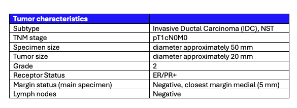 Table 1: Tumor and lymph node characteristics assessed by histopathological evaluation.
Histopathological evaluation
After PET-CT imaging, the surgical specimen was sent to the pathology department for routine histopathological evaluation, which was available after several days. The histopathological results are listed in Table 1.
Surgical margins were tumor-free, with a minimum margin of 5 mm medially. The margin towards the skin was 0.5 mm. Histopathology results of the sentinel node confirmed no metastatic deposit was present.
Discussion and conclusion
This case underscores the value of integrating specimen PET-CT imaging into the operating room, providing surgeons with real-time confidence in their decisions. Moreover, the initial goal was to preserve the skin. However, during surgery, the surgeon palpated the tumor, which was extending up to the skin border. For this reason, the surgeon opted to remove the overlying portion of skin. This clinical assessment aligned with the specimen PET-CT images, which showed 18F-FDG uptake approaching—but not reaching—the skin area. This confirmation reassured the surgeon that her decision was correct, and that the entire tumor had been successfully resected. Final histopathology confirmed that tumor cells almost reached the border of the skin area, and that final margins were negative.
References
[1] St John et al. (2017). Diagnostic Accuracy of Intraoperative Techniques for Margin Assessment in Breast Cancer Surgery: A Meta-analysis. Ann Surg.
[2] Göker M. et al. (2020). 18F-FDG micro-PET/CT for intra-operative margin assessment during breast-conserving surgery. Acta Chirurgica Belgica. https://doi.org/10.1080/00015458.2020.1774163
[3] Lambert B. et al. (2025) Feasibility study on the implementation of a mobile high-resolution PET/CT scanner for surgical specimens: exploring clinical applications and practical considerations. Eur J Nucl Med Mol Imaging. https://doi.org/10.1007/s00259-025-07143-z