Imaging Case 3:
Breast - Ductal carcinoma in situ
A 63-year-old woman diagnosed with ductal carcinoma in situ underwent wire-guided breast-conserving surgery.
 Images and case information courtesy of dr. Menekse Göker, Department of Gynaecology, Ghent University Hospital.”
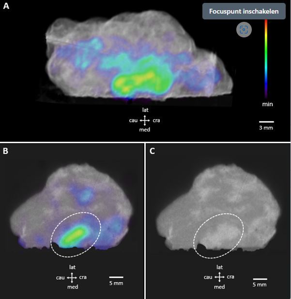
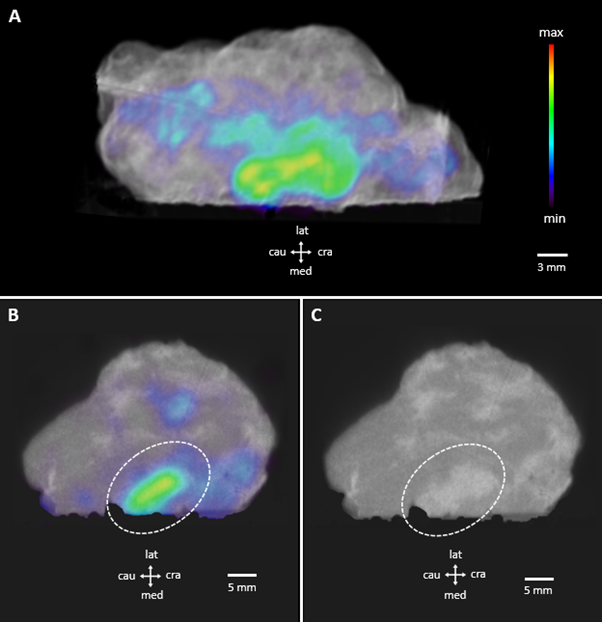
The patient was intravenously injected with 18F-FDG prior to breast-conserving surgery at the nuclear medicine department. Figure 1A shows a 3-dimensional render of the PET-CT images of the resected ductal carcinoma in situ. The 18F-FDG distribution is represented in color scale superimposed on the CT render in greyscale. A main hotspot of 18F-FDG uptake is visible at the medial side (lower edge of the image) of the resected breast tissue. Figure 1B and Figure 1C show a cross section of the PET-CT images and the CT only images respectively. The dashed ellipse on Figure 1B indicates that 18F-FDG is visible at the specimen border. The CT also confirms that there is dense tissue reaching the tissue border (Figure 1C, dashed ellipse).
Figure 1 demonstrates PET-CT specimen images of a resected ductal carcinoma in situ. A 3-dimensional PET-CT render (A), a PET-CT cross section (B) and a CT only cross section (C) are illustrated. The dashed ellips indicates a suspicious margin at the medial side of the resected breast tissue. Specimen orientation is as indicated.
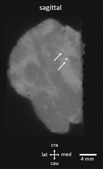
Microcalcifications can be detected on the CT images. Figure 2 shows the sagittal section of the CT scan. The white arrows indicate the microcalcifications located inside the ductal carcinoma in situ. In addition to the information provided by the PET image, this may also allow confirming that all microcalcifications were resected.
Figure 2 shows sagittal view of the CT scan of the resected ductal carcinoma in situ. Microcalcifications can be clearly observed on the CT images (white arrows).