Imaging Case 34
Head & Neck Cancer - Right Oropharyngeal Squamous Cell Carcinoma
Achieving complete tumor resection with adequate margins is fundamental in head and neck oncology surgery, as the resection status is a strong predictor of overall survival and recurrence-free survival. Intraoperative margin assessment is therefore essential yet remains challenging in anatomically complex regions such as the oropharynx, where treatment for squamous cell carcinoma aims for a minimal marginal clearance of 5 mm. Currently, frozen section analysis is the standard method for intraoperative margin assessment. Frozen sections of the tumor bed can identify positive margins but cannot reliably detect close margins (<5 mm). Moreover, false-negative findings may leave residual disease undetected, while false-positive findings can lead to unnecessary additional resection and surgical overtreatment.
Intraoperative specimen PET-CT imaging has been investigated as an additional method to assess resection margins directly in the operating room. By imaging the resected specimen, this approach may provide real-time information on tumor extent and margin proximity, complementing intraoperative pathology.
This case is presented with the support of Dr. med. Kazim Shiraliyev, Prof. Dr. med. Moritz Meyer, PD Dr. med. Anke Daser and Dr. med. Eric Deuß of the University Hospital Essen - ENT Clinic, Germany.
Patient History
A 75-year-old male patient was diagnosed with a right-sided oropharyngeal carcinoma. Preoperative histology identified a poorly differentiated, p16 positive squamous cell carcinoma. Clinical staging was cT2 N0 M0.
Following multidisciplinary discussion, the team and the patient opted for surgical treatment. Shortly, a right-sided partial oropharyngeal resection with bilateral neck dissection was performed with curative intent.
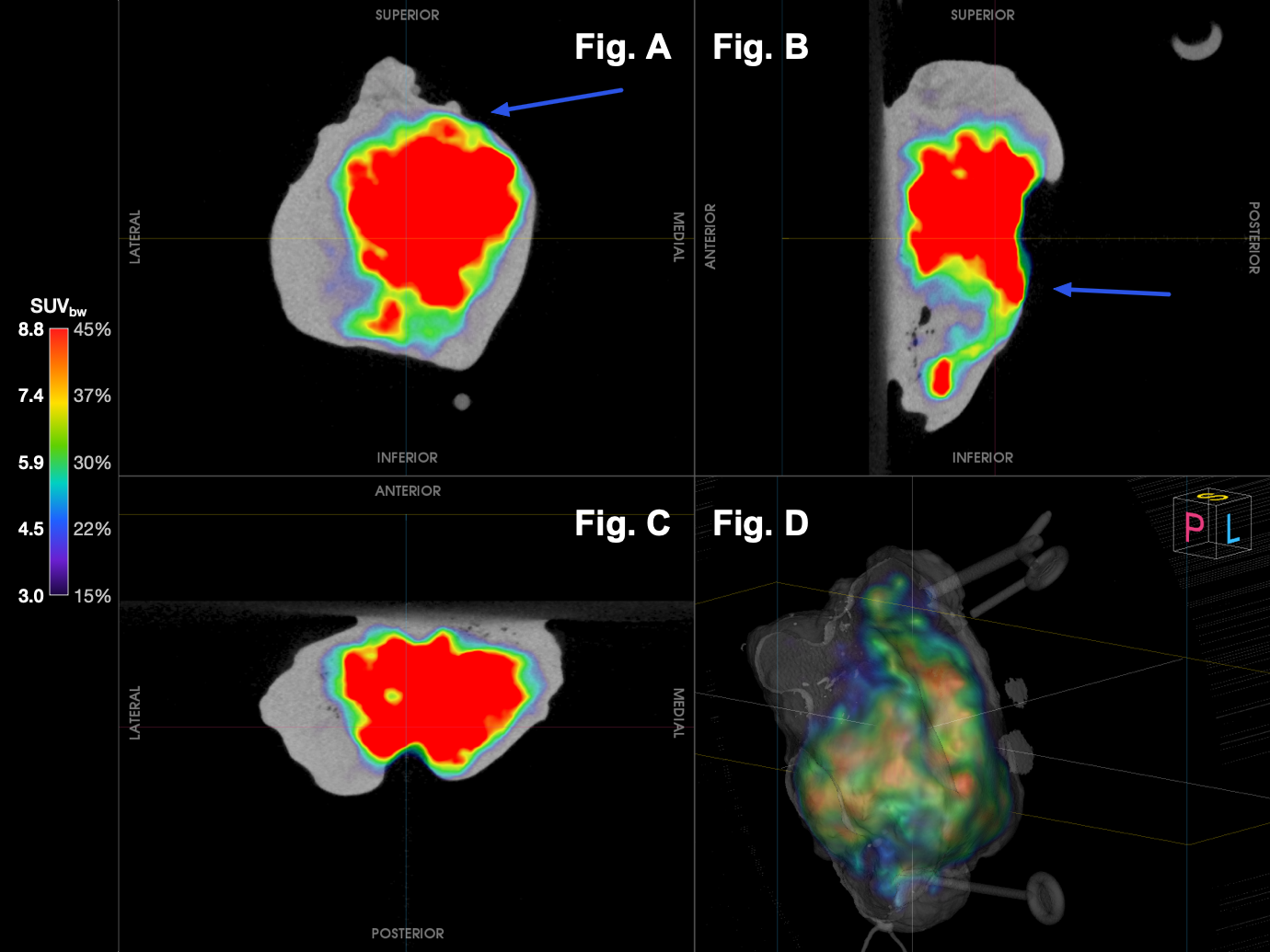
[Fig. 1] Intraoperative specimen PET-CT of the freshly resected right oropharyngeal/tonsillar specimen, acquired on the AURA 10 PET-CT. A) Coronal aspect. B) Sagittal aspect. C) Axial/transversal aspect. D) 3D PET-CT rendering. The colour scale encodes ¹⁸F-FDG activity concentration from 3.0 to 8.8 SUVbw (15–45% of maximum). Intense tracer uptake fills most of the specimen and extends toward the superior-medial aspect, where it reaches closely to the specimen edge with significant uptake; this corresponds to the close cranial deep margin (1 mm) on definitive histology, which prompted the intraoperative cavity shave. Uptake reaching the posterior aspect corresponds to the mucosal (luminal) surface of the oropharynx, i.e. a free epithelial/air interface rather than a true resection margin and should not be interpreted as margin involvement.
Specimen PET-CT Imaging
cranial tumor bed during the same procedure.
Histopathological Evaluation
Postoperative histological evaluation of the main specimen demonstrated a p16-positive (HPV-associated) squamous cell carcinoma of the right oropharynx with pathological staging pT2N0 (0/60) L0 V0 Pn0.
Definitive histology of the main specimen confirmed an R0 resection, with tumor approaching but not reaching the cranial deep margin. The minimum clearance at this margin was 1 mm, i.e. a close margin.
A pathology summary described a formalin-fixed right tonsillar specimen measuring 4.6 x 3.2 x 1.9 cm, containing a poorly defined firm grey-white tumor measuring 2.9 x 2.3 x 1.6 cm. The minimum distances to the resection margins were reported as 3 mm at 12 o'clock, 3 mm at 3 o'clock, 4 mm at 6 o'clock, 2 mm at 9 o'clock and 1 mm at the basal (cranial deep) margin.
Although the intraoperative frozen sections of the tumor bed were tumor-free and the main specimen was R0, the cranial deep margin was close (1 mm). Because intraoperative specimen PET-CT has shown an accumulation of ¹⁸F-FDG reaching the margin of the specimen, a cavity shave was taken from the corresponding tumor bed during the primary intervention. Final pathology confirmed the shave was tumor-free, securing adequate clearance at the cranial margin in a single procedure.
At 6 months postoperatively, follow-up ¹⁸F-FDG PET-CT showed only physiological and post-surgical activity at the resection site, with no evidence of residual or recurrent disease (Figure 2).
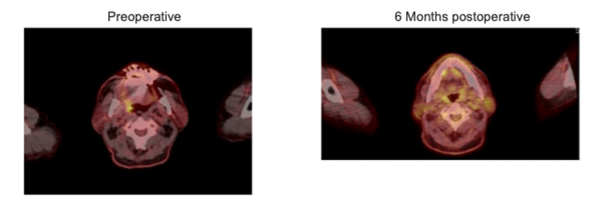 [Fig. 2] Diagnostic ¹⁸F-FDG PET-CT, transverse fused images at the level of the oropharynx. Left (preoperative): focal FDG-avid lesion in the right oropharynx/tonsillar region corresponding to the primary p16-positive cT2 N0 carcinoma. Right (6 months postoperative): no abnormal focal uptake at the resection site, with only physiological/post-surgical activity, consistent with the adequate clearance at the cranial margin after cavity shaving .
Discussion and Conclusion
This case demonstrates the unique and complementary role of intraoperative specimen PET-CT in margin assessment during
head and neck surgery.
Intraoperative frozen sections of the tumor bed were disease-free and failed to identify a margin at risk. While final pathology confirmed the main specimen was R0 with a close, 1 mm cranial deep margin. Specimen PET-CT was capable of visualizing tumor reaching close to this margin, providing crucial information that the frozen sections did not capture. The clinical significance of specimen PET-CT lies in its ability to directly influence intraoperative decision making. Despite negative frozen sections, the surgical team performed an immediate additional resection during the primary procedure, as the specimen PET-CT findings provided complementary evidence supporting the surgical decision. This suggests that the intraoperative decision prompted by specimen PET-CT allowed achievement of a secure and complete resection. The clean 6-month follow-up provides early evidence that this intraoperative intervention translated into a durable clinical benefit for the patient.
By providing immediate intraoperative feedback, specimen PET-CT imaging demonstrates its added value as a complementary tool for intraoperative margin assessment and surgical decision making. This results in more confidence and potentially improved oncologic outcomes.