Story
Imaging Case 35:
Head & Neck Cancer - Floor of Mouth Squamous Cell Carcinoma
Achieving adequate resection margins of at least 5 mm remains a key challenge in head and neck oncology, particularly in anatomically complex regions such as the floor of the mouth. Close or involved margins are associated with an increased risk of recurrence and often necessitate adjuvant therapy, which can lead to significant morbidity and long-term functionalconsequences for patients.[1]
In addition, final histopathological evaluation typically requires a minimum of 7–10 days, limiting the opportunity to take corrective action during the primary surgical procedure. In this context, intraoperative specimen PET-CT imaging has the potential to provide real-time insight into tumor extent and margin status, directly supporting surgical decision-making at
the point of care.[2]
Patient History & Surgical Procedure
A 50-year-old male patient was diagnosed with advanced squamous cell carcinoma of the floor of the mouth. Preoperative staging indicated cT4a N3b disease, with evidence of extensive locoregional involvement.The patient underwent a composite resection including mandibulectomy and bilateral neck dissection at a large academic medical center in the United States. The bilateral neck dissection consisted of a right selective neck dissection of levels I–IV and a left modified radical neck dissection of levels I–IV. Reconstruction was achieved using a fibular free flap.
During the procedure, two tumor bed-based margins (the left submandibular gland nerve margin and the right mandibular margin) were assessed by intraoperative frozen section analysis. Despite initial concern regarding possible deep tumor extension toward the neck, all results were negative for tumor involvement.
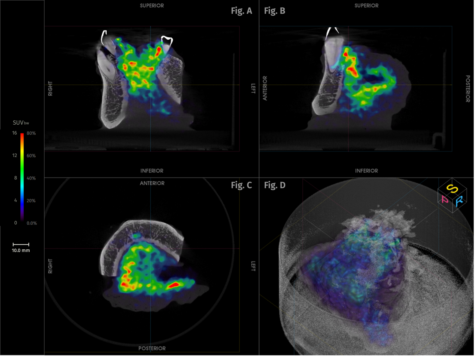
[Fig. 1] High-resolution intraoperative PET-CT of the resected specimen — mandible, floor of mouth and tongue — acquired on the AURA 10 PET-CT. SUVbw scale 0 – 16. A) Coronal aspect. B) Sagittal aspect. C) Axial/transversal aspect. D) 3D PET-CT rendering.
Specimen PET-CT Imaging
During surgery, the patient received an injection of 0.8 MBq/kg
(equivalent to 0.022 mCi/kg) of ¹⁸F-FDG. Four hours after injection, the tumor resection was completed. Immediately following resection, high-resolution specimen PET-CT imaging was performed intraoperatively. Within 10 minutes, the intraoperative specimen imaging on the AURA 10 PET-CT was available.
Based on the PET-CT images, the tumor clearly extended into the base of the tongue and the anterior mandible bone. There was no indication of tumor involvement at the resection margins, supporting the intraoperative assessment of complete tumor removal.
Histopathological Evaluation
Final histopathological evaluation confirmed a moderately differentiated squamous cell carcinoma measuring approximately 3.5 cm, located in the anterior floor of the mouth. The tumor showed extensive local invasion, involving the floor of the mouth, the base of the tongue, the submandibular gland, and extending through the full thickness of the mandible.
All resection margins were negative for invasive tumor, with the closest margins measuring 2 mm at the superior and posterior soft tissue margins, which was highly consistent with distances measured on the intraoperative AURA 10 PET-CT images.
Extensive regional lymph node involvement was observed bilaterally, with metastatic disease present across multiple nodal levels. Extranodal extension was identified, indicating aggressive nodal disease. Based on these findings, the final pathological staging was pT4a N3b.
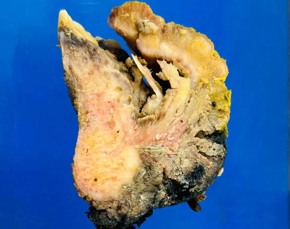 [Fig. 2] Gross pathology specimen: sagittal section through the composite resection including mandible, floor of mouth and tongue.
Discussion and Conclusion
This case illustrates the unique value of intraoperative specimen PET-CT imaging in the surgical management of advanced head and neck cancer. The imaging provided real-time information supporting the assessment of tumor extent and margin status.Importantly, the findings from specimen PET-CT imaging were consistent with both frozen section analysis and final histopathological evaluation, particularly with respect to margin status and local invasion.
By providing immediate feedback on resection completeness, specimen PET-CT imaging may enhance surgical confidence and support intraoperative decision-making in a complex oncological resection. Ultimately, this approach has the potential to optimize surgical outcomes and reduce the need for additional treatment in selected patients.
References
1. Machiels JP, Leemans CR, Golusinski W, et al. Squamous cell carcinoma of the oral cavity, larynx, oropharynx and hypopharynx: EHNS–ESMO–ESTRO Clinical Practice Guidelines. Ann Oncol. 2020;31(11):1462-75.2. Lambert B, et al. Feasibility study on the implementation of a mobile high-resolution PET/CT scanner for surgical specimens. Eur J Nucl Med Mol Imaging. 2025. doi:10.1007/s00259-025-07143-z