Imaging Case 36:
Head & Neck Cancer - Recurrent Soft Palate Squamous cell Carcinoma
Achieving complete tumor resection with adequate margins remains a key objective in head and neck oncology. This is particularly challenging in cases of recurrent disease, where prior surgeries may alter anatomy and tumor behavior. Recurrent tumors may demonstrate irregular growth patterns and subclinical extension, complicating intraoperative assessment of the tumor extent.
Intraoperative specimen PET-CT imaging enables real-time visualization of metabolic tumor activity within the resected specimen and may support the evaluation of tumor localization and margin status during surgery.
Patient History
A 65-year old female patient presented with a history of recurrent squamous cell carcinoma of the soft palate. The patient had
previously undergone multiple resections for palate and tongue tumors, each time achieving negative margins, yet with continue tumor recurrence over time. At presentation, the patient was diagnosed with recurrent soft palate squamous cell carcinoma, clinically staged as cT3N1M0.
Surgical management was undertaken at a large academic medical center in the United States and consisted of radical resection of the soft palate lesion. Notably, no neck dissection was performed despite clinical suspicion of nodal disease. Following induction of anesthesia, the patient received an intravenous injection of 0,8 MBq/kg (equivalent of 0,022 mCi/kg)¹⁸F-FDG as part of a clinical trial.
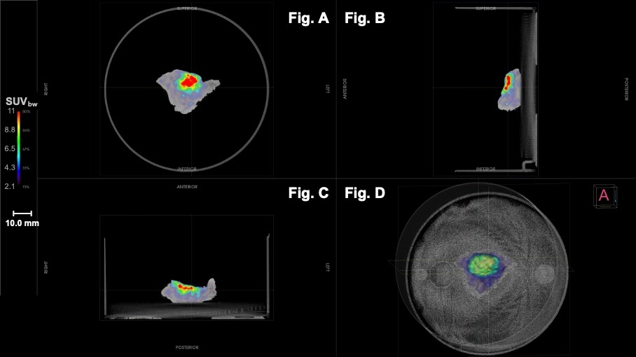
Figure 1: High-resolution PET-CT images of the resected soft palatespecimen. A) Coronal aspect. B) Sagittal aspect; ¹⁸F-FDG uptake
reaches the anterior border. C) Axial/transversal aspect;¹⁸F-FDG
uptake reaches the anterior border. D) 3D PET-CT image of the
specimen, including the base of the specimen container.
Specimen PET-CT Imaging
Intraoperative specimen PET-CT imaging was performed immediately following resection. Within 10 minutes the intraoperative specimen PET-CT imaging demonstrated focal radiotracer uptake corresponding to the tumor. An area of increased uptake was observed at the anterior aspect of the specimen; as this margin is oriented towards the oral cavity, it was deemed not clinically relevant. Furthermore, three-dimensional orientation of the tumor provided by the AURA 10 PET-CT enabled the surgeon to assess the distance to the superior margin more accurately. No additional suspicious radiotracer uptake was observed extending to the specimen margins.
Intraoperative Decision Making
Following specimen PET-CT imaging, the specimen was submitted for frozen section analysis to assess margin status. Frozen sections demonstrated that all assessed margins, including deep, superior, and inferior/lateral margins, were negative for
carcinoma and dysplasia. Based on the combined imaging findings and frozen section results, no additional resection was performed, and the procedure was concluded.
Histopathological Evaluation
Final histopathological evaluation confirmed a moderately differentiated keratinizing squamous cell carcinoma of the left soft
palate, measuring 2.5 cm in greatest dimension. All surgical margins were negative for invasive carcinoma with the closest margin measuring 4 mm at the deep margin, which was highly concordant with the corresponding intraoperative AURA 10 PET-CT images. The tumor was staged as pT2 according to the AJCC 8th edition staging system.
Discussion and Conclusion
This case highlights the potential value of intraoperative specimen PET-CT imaging in the surgical management of recurrent head and neck cancer. Specimen PET-CT imaging enabled real-time visualization of tumor metabolic activity and supported intraoperative assessment of tumor localization. Increased uptake at the anterior aspect of the specimen corresponded to the mucosal surface of the oral cavity rather than a true surgical margin, and was not considered clinically relevant.
The imaging findings were consistent with both frozen section analysis and final histopathological evaluation, all of which confirmed complete tumor resection with negative margins. In the setting of recurrent disease, where surgical decision-making is inherently more complex, specimen PET-CT imaging may offer valuable real-time information to support intraoperative assessment while helping to avoid unnecessary extension of resection.
References
1. Hamoir M, et al. The current role of salvage surgery in recurrent head and neck squamous cell carcinoma. Cancers (Basel). 2018;10(8):267.
2. Hartl DM, et al. Review of outcomes after salvage surgery for recurrent squamous cell carcinoma of the head and neck.
Cancers (Basel). 2023;15(19):4692.
3. Lambert B, et al. Feasibility study on the implementation of a mobile high-resolution PET/CT scanner for surgical specimens.
Eur J Nucl Med Mol Imaging. 2025. doi: 10.1007/s00259-025-07143-z.