Imaging Case 4:
Breast Cancer - Invasive Ductal Carcinoma
During breast-conserving surgery it is common practice to perform intraoperative assessment of the resected tumor specimen to assess if the resection was successful. The most frequently used techniques such as specimen radiography or frozen section assessment all have their drawbacks regarding performance and/or efficiency [1].
In this clinical case, which is part of the multi-center prospective BrIMA study (NCT04999917), we highlight how intraoperative specimen PET-CT imaging can bring more confidence into the operating room. This gives the surgeon a clear view of what was resected – and the ability to act upon this information at the point of surgery.
Patient history
A 63-year-old woman with invasive ductal carcinoma underwent breast-conserving surgery followed by sentinel node resection. The tumor size on preoperative imaging was 14mm, biopsy showed a grade 1 tumor with receptor status ER+/PR+/HER2-. Preoperative staging was cT1cN0M0.PET-CT Specimen images
The patient received a periareolar injection with 158 MBq of 99mTc and an intravenous injection with 53 MBq (0.80 MBq/kg) of 18F-FDG at the nuclear medicine department. The patient was then transferred to the operating theatre and breast-conserving surgery was performed per standard protocol.The resected breast specimen was imaged immediately after resection and the images were interpreted by the surgeon in the operating room. This was approximately 90 min after 18F-FDG injection.
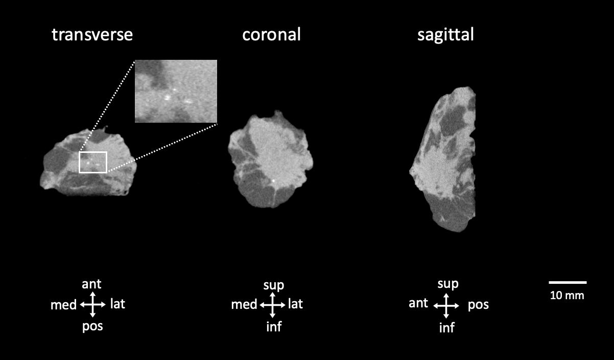 Figure 1. Transverse, coronal and sagittal slices of the CT specimen images of an invasive ductal carcinoma. A dense lump is visualized slightly excentric in the specimen towards the antero-infero-lateral border of the specimen. Microcalcifications can be seen (inset). Abbreviations: med, medial; lat, lateral; sup, superior; inf, inferior.
The CT (figure 1) visualizes the entire specimen in 3D and shows a dense lump in the specimen, localized slightly towards the inferolateral side of the specimen. This already provides an indication of where the tumor is located in the specimen. Several microcalcifications can be observed due to the high spatial resolution (100µm) of the CT image.
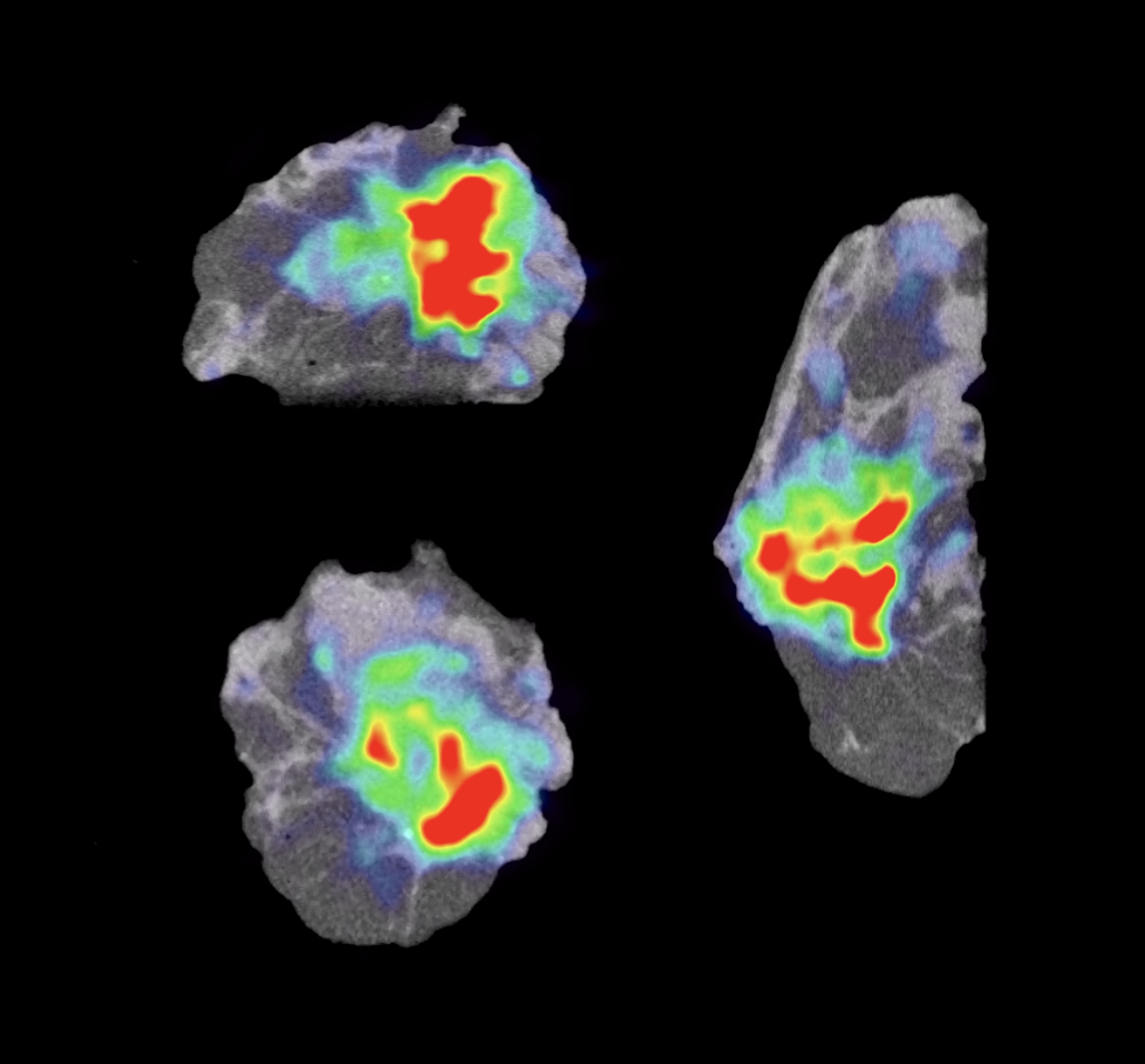
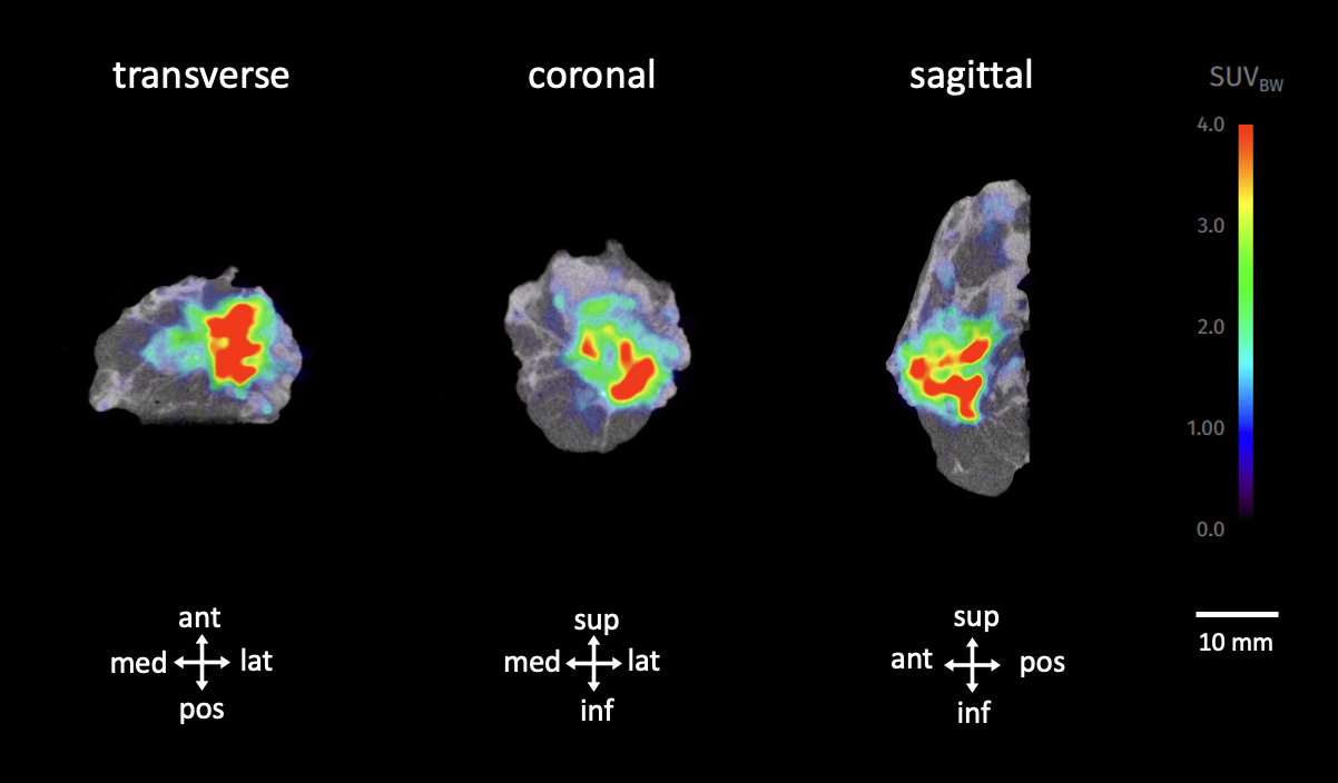 Figure 2. Transverse, coronal and sagittal slices of the PET-CT specimen images of an invasive ductal carcinoma. The 18F-FDG uptake is visible in the color overlay and reaches the antero-infero-lateral border of the specimen. Final histopathology confirmed a positive surgical margin inferolateral. Specimen orientation is as indicated. Abbreviations: med, medial; lat, lateral; sup, superior; inf, inferior.
Adding the PET overlay to the CT images demonstrates how specimen PET-CT leverages to power of molecular imaging to bring more clarity into the operating room (figure 2). The colored overlay demonstrates increased 18F-FDG uptake, which indicates metabolically more active regions such as invasive ductal carcinoma cells.
In this case,18F-FDG uptake reaches the antero-infero-lateral border of the imaged specimen. Based on this image, the surgeon decided to take a cavity shave in this area. Postoperative final pathology showed that there was indeed a positive margin in this area
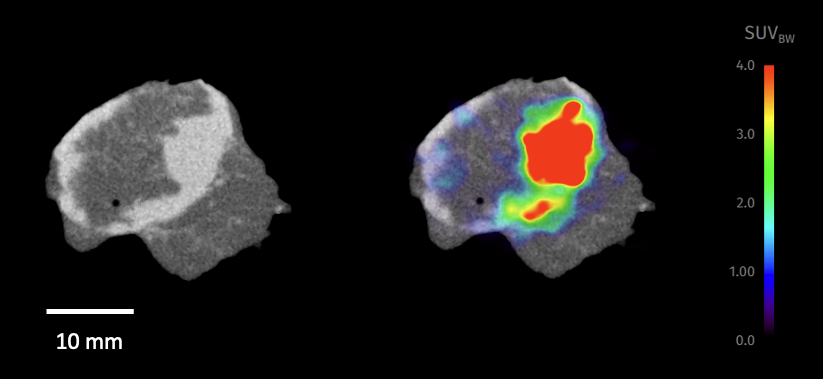 Figure 3. CT (left) and PET-CT (right) image of the sentinel lymph node (left). The dense (light grey) circular structure represents the resected lymph node. On the PET image a clear region of 18F-FDG uptake is visualized in the lymph node. Final histopathology confirmed metastatic invasion of the node.
After tumorectomy the sentinel lymph node was localized using a gamma probe and resected. A specimen PET-CT image was also acquired of the sentinel node (figure 3). The CT images visualize a circular dense structure allowing confirmation that a lymph node was resected. The PET images show increased 18F-FDG uptake in the node, leading to the suspicion of malignant invasion of the sentinel node. A metastatic deposit of 12mm was shown by postoperative final pathology.
Histopathological evaluation
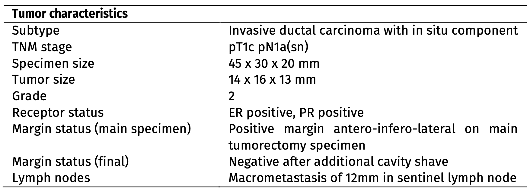
After PET-CT specimen imaging, the resected specimens were sent to the pathology department for routine histopathological evaluation. The final histopathological results are presented in Table 1. The final margin status – after the additional cavity shave – was negative. No reoperation was required and the patient could immediately move on to postoperative therapy.
Discussion and conclusion
This case highlights the power of bringing specimen PET-CT imaging into the operating room. The surgeon decided intraoperatively to perform an additional cavity shave as imaging indicated 18F-FDG uptake was reaching the border of the specimen. Final histopathology confirmed that tumor cells reached the border of the specimen in this area. Taking the extra cavity shave possibly avoided the need for reoperation and the associated burden in this patient.
REFERENCES
[1] St John et al, Diagnostic Accuracy of Intraoperative Techniques for Margin Assessment in Breast Cancer Surgery: A Meta-analysis. Ann Surg 2017.