Imaging Case 6:
Head & Neck - Squamous Cell Carcinoma of the scalp
Cutaneous squamous cell carcinoma (cSCC) is the second most common non-melanoma skin cancer. They arise commonly in the head and neck region, especially on the scalp. For most patients complete tumor resection with an adequate margin of healthy tissue (> 5mm) is the first line of treatment. This case illustrates how specimen PET-CT images could help the surgeon intraoperatively remove the target lesion. It was published as part of a pilot study in cancer of the head and neck region.
Patient History
A 78-year-old man was scheduled for a resection of a squamous cell carcinoma (SCC) of the scalp.
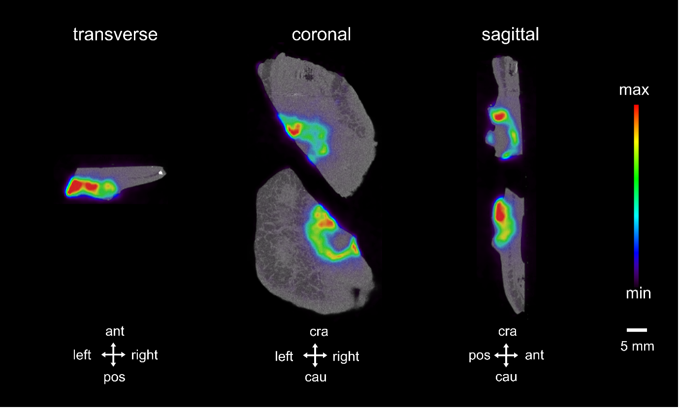 Figure 1: transverse, coronal and sagittal slides of the PET-CT scan of two halves of a scalp specimen. A central 18F-FDG hotspot is visible. Surgical orientation of the specimen images is shown at the bottom of each slice. The PET tracer scale bar is depicted at the right hand side. Abbreviations: ant, anterior; pos, posterior; cra, cranial; cau, caudal.
PET-CT specimen images
On the day of surgery, 18F-FDG was intravenously administered to the patient right before the patient was put under anesthesia to start the surgery. The tumor was surgically removed and a PET-CT image of the surgical specimen was acquired immediately after resection of the specimen. The specimen was cut by a pathologist in two halves before performing the PET-CT scan.
Figure 1 shows three orthogonal views of the PET-CT specimen images. The PET images are represented in color scale superimposed on the CT images in greyscale. A circular hotspot of 18F-FDG uptake is visible in the centre of the specimen.
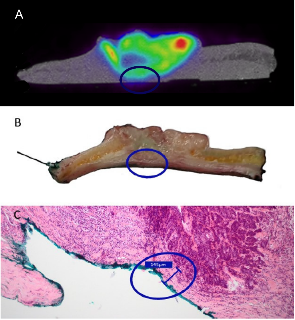
 Figure 2: correlation between the close margin as seen on a transverse view of the PET-CT images (A), on a slice of the resected tissue (B) and on a close-up of a whole-slide pathology image (C). The region of interest with the close margin is indicated with a blue circle.
Findings
After PET-CT imaging, the surgical specimen was sent to the pathology department for routine histopathological evaluation. A pathological close margin between the invasive tumor and the resection margin of 145 µm was found. When correlating the PET-CT images with the pathology whole-slide images, this close margin could be located on the PET-CT images as a small region where the deep margin shows an increased 18F-FDG uptake. This is visualized in Figure 2.
Discussion and conclusion
When correlating the PET-CT images with the whole-slide pathology images, a good correspondence was found between 18F-FDG activity close to the resection margin in the PET-CT images and the position of a close margin as confirmed after histopathological examination. Having this information at hand during surgery could help the surgeon decide to extend the resection to ensure an adequate free margin.
REFERENCES
Rogers HW et al. Incidence estimate of nonmelanoma skin cancer (keratinocyte carcinomas) in the US population, 2012. JAMA Dermatol 2015; 151(10): 1081-1086.
Alam M et al. Guidelines of care for the management of cutaneous squamous cell carcinoma. J Am Acad Dermaol 2018; 78(3): 560-578.
Debacker JM et al. High-Resolution 18F-FDG PET/CT for Assessing Three-Dimensional Intraoperative Margins Status in Malignancies of the Head and Neck, a Proof-of-Concept. J Clin Med 2021; 10:3737.