Imaging Case 8:
Pancreatic adenocarcinoma
Pancreatic adenocarcinoma is the most frequent tumor occurring in the pancreas. Early-stage pancreatic cancers can be treated surgically, but there is a high-rate of positive surgical margins, which may be associated with a worse prognosis. Therefore, intraoperative evaluation of the resected specimen to ensure free surgical margins could be of interest.
In this clinical case we show how a detailed view of the resected pancreas specimen can be obtained with high-resolution specimen PET-CT imaging. The information and images in this case stem from a pilot study performed by prof. Frederik Berrevoet and his team at Ghent University Hospital.
Patient History and preparation
A patient was diagnosed with a poorly differentiated ductal adenocarcinoma of the pancreas and was scheduled for a pylorus-sparing Whipple resection. On the day of surgery, 274 MBq of 18F-FDG was injected in the operating room at the start of surgery.
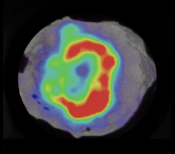
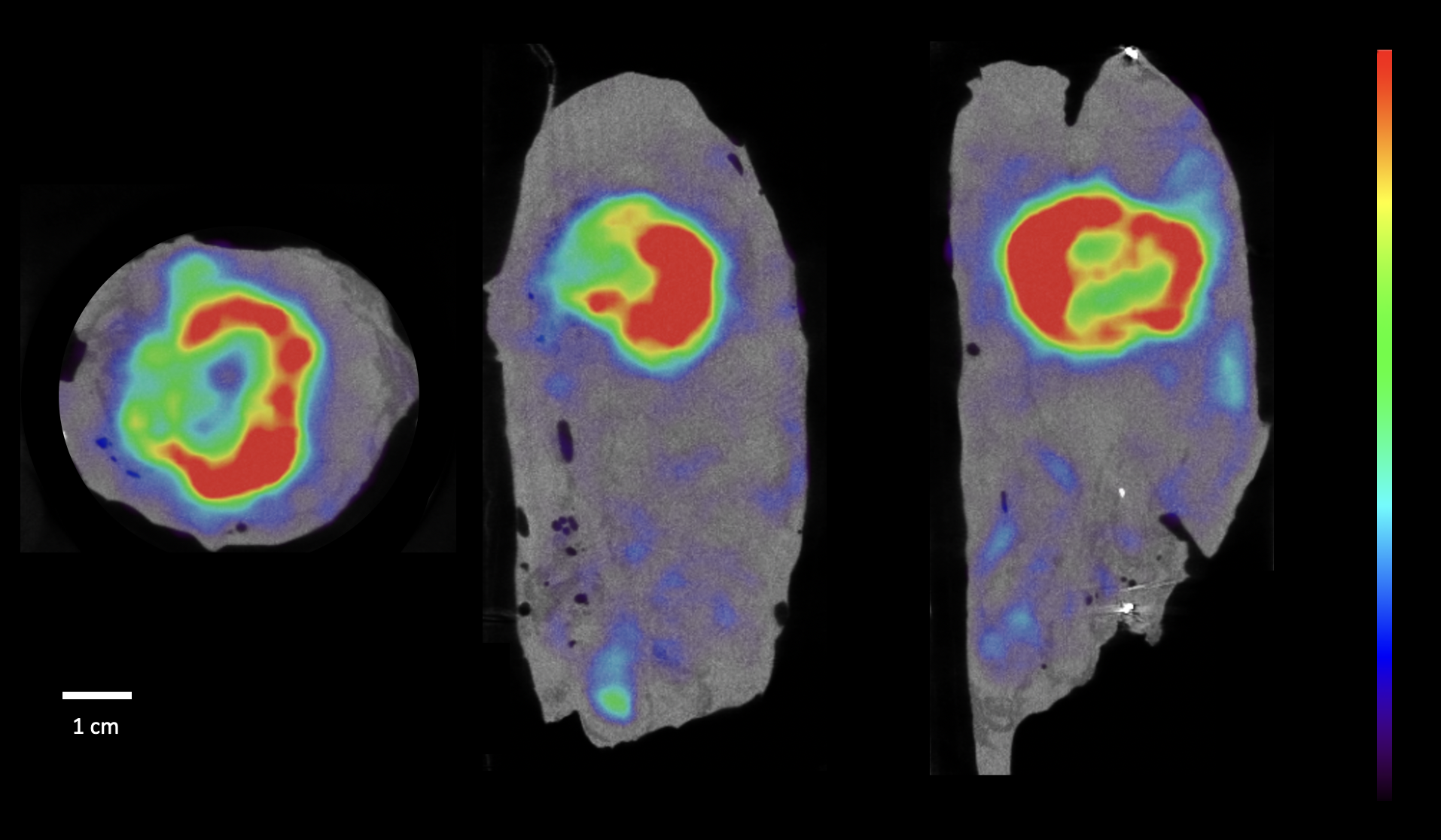 Figure 1: high-resolution specimen PET-CT image of the resected pancreas specimen. A distinct region of heterogenous 18F-FDG uptake is visualized in the caput.
PET-CT specimen images
After resection of the specimen, a high-resolution PET-CT image was acquired. The time between radiotracer injection and acquisition was 335 minutes (approximately 3 half-lives of 18F). Fig. 1 shows three orthogonal slices of the specimen PET-CT image. A distinct region of heterogeneous 18F-FDG uptake, which does not reach the border of the specimen, is visible in the caput of the pancreas.
Histopathology
Histopathology confirmed a tumor of 3.6 x 2.5 x 2 cm in the caput of the pancreas. The resection margins were free of tumor cells (closest margin = 1.2mm). There was lymphovascular and perineural invasion present. Postoperative staging was pT2N2.
Discussion and conclusion
This case illustrates how specimen PET-CT imaging can be used to visualize pancreatic adenocarcinoma with high spatial resolution. The PET-CT images demonstrate a well-delineated region of uptake that does not reach the border of the specimen. This was correlated to histopathology, where the resection margins were deemed free.
REFERENCES
T. Nitta et al. The impact of margin status determined by the one-millimeter rule on tumor recurrence and survival following pancreaticoduodenectomy for pancreatic ductal adenocarcinoma. Surg Today. 2017 Apr;47(4):490-497.
L. Maris et al. 18F-FDG-PET/CT specimen imaging for perioperative visualization of pancreatic adenocarcinoma: a proof-of-concept study. 41st congress of the European Society of Surgical Oncology Abstracts, 2022.